From Surf Wiki (app.surf) — the open knowledge base
Striatum
Nucleus in the basal ganglia of the brain
Nucleus in the basal ganglia of the brain
| Field | Value |
|---|---|
| Name | Striatum |
| Latin | striatum |
| Image | Cortical surface with an overlay of the basal ganglia and thalamus.jpg |
| Caption | Striatum shown in green with other basal ganglia and thalamus. Small region in yellow is the amygdala |
| Image2 | Corticostriatal Pathway.jpg |
| Caption2 | Tractography showing corticostriatal connections |
| IsPartOf | Basal ganglia |
| Reward system | |
| Components | Ventral striatum |
| Dorsal striatum |
Reward system Dorsal striatum
The striatum (: striata) or corpus striatum is a cluster of interconnected nuclei that make up the largest structure of the subcortical basal ganglia. The striatum is a critical component of the motor and reward systems; receives glutamatergic and dopaminergic inputs from different sources; and serves as the primary input to the rest of the basal ganglia.
Functionally, the striatum coordinates multiple aspects of cognition, including both motor and action planning, decision-making, motivation, reinforcement, and reward perception. The lentiform nucleus is made up of the larger putamen, and the smaller globus pallidus. Strictly speaking the globus pallidus is part of the striatum. It is common practice, however, to implicitly exclude the globus pallidus when referring to striatal structures.
In primates, the striatum is divided into the ventral striatum and the dorsal striatum, subdivisions that are based upon function and connections. The ventral striatum consists of the nucleus accumbens and the olfactory tubercle. The dorsal striatum consists of the caudate nucleus and the putamen. A white matter nerve tract (the internal capsule) in the dorsal striatum separates the caudate nucleus and the putamen.
Structure
The striatum is the largest structure of the basal ganglia. The striatum is divided into two subdivisions, a ventral striatum and a dorsal striatum, based upon function and connections. It is also divisible into a matrix and embedded striosomes.
Ventral striatum
The ventral striatum is composed of the nucleus accumbens and the olfactory tubercle. The nucleus accumbens is made up of the nucleus accumbens core and the nucleus accumbens shell, which differ by neural populations. The olfactory tubercle receives input from the olfactory bulb but has not been shown to play a role in processing smell. In non-primate species, the islands of Calleja are included. The ventral striatum is associated with the limbic system and has been implicated as a vital part of the circuitry for decision making and reward-related behavior.
Dorsal striatum
The dorsal striatum is composed of the caudate nucleus and the putamen. Primarily it mediates cognition and involves motor and executive function. The dorsal striatum can be further subdivided into the dorsomedial striatum, and the dorsolateral striatum. Both of these areas have different roles in the acquisition of learnt behaviour and skill formation. The dorsomedial region receives projections from the frontal and the parietal cortices. The dorsolateral region receives projections from the sensorimotor cortex.
Matrix and striosomes
Neurochemistry studies have used staining techniques on the striatum that have identified two distinct striatal compartments, the matrix, and the striosome (or patch). The matrix is seen to be rich in acetylcholinesterase, while the embedded striosomes are acetylcholinesterase-poor. The matrix forms the bulk of the striatum, and receives input from most areas of the cerebral cortex. Clusters of neurons in the matrix, called matrisomes receive a similar input. Their output goes to both regions of the globus pallidus and to the substantia nigra pars reticulata.
The striosomes receive input from the prefrontal cortex and give outputs to the substantia nigra pars compacta. There are more striosomes present in the dorsal striatum making up 10-15% of the striatal volume, than in the ventral striatum.
Cell types
Types of cells in the striatum include:
- Medium spiny neurons (MSNs), which are the principal neurons of the striatum.
- Cholinergic interneurons release acetylcholine, which has a variety of important effects in the striatum. In humans, other primates, and rodents, these interneurons respond to salient environmental stimuli with stereotyped responses that are temporally aligned with the responses of dopaminergic neurons of the substantia nigra. The large aspiny cholinergic interneurons themselves are affected by dopamine through D5 dopamine receptors. Dopamine also directly controls communication between cholinergic interneurons.
- There are many types of GABAergic interneurons. The best known are parvalbumin expressing interneurons, also known as fast-spiking interneurons, which participate in powerful feedforward inhibition of principal neurons. Also, there are GABAergic interneurons that express tyrosine hydroxylase, somatostatin, nitric oxide synthase and neuropeptide-y. Recently, two types of neuropeptide-y expressing GABAergic interneurons have been described in detail, one of which translates synchronous activity of cholinergic interneurons into inhibition of principal neurons. These neurons of the striatum are not distributed evenly.
There are two regions of neurogenesis in the brain – the subventricular zone (SVZ) in the lateral ventricles, and the dentate gyrus in the hippocampal formation. Neuroblasts that form in the lateral ventricle adjacent to the striatum, integrate in the striatum. This has been noted in the human striatum following an ischemic stroke. Injury caused to the striatum stimulates the migration of neuroblasts from the SVZ, to the striatum, where they differentiate into adult neurons. The normal passage of SVZ neuroblasts is to the olfactory bulb but this traffic is diverted to the striatum after an ischemic stroke. However, few of the new developed neurons survive.
Inputs
The largest connection is from the cortex, in terms of cell axons. Many parts of the neocortex innervate the dorsal striatum. The cortical pyramidal neurons projecting to the striatum are located in layers II-VI, with the most dense projections come from layer V. They end mainly on the dendritic spines of the spiny neurons. They are glutamatergic, exciting striatal neurons.
The striatum is seen as having its own internal microcircuitry. The ventral striatum receives direct input from multiple regions in the cerebral cortex and limbic structures such as the amygdala, thalamus, and hippocampus, as well as the entorhinal cortex and the inferior temporal gyrus. Its primary input is to the basal ganglia system. Additionally, the mesolimbic pathway projects from the ventral tegmental area to the nucleus accumbens of the ventral striatum.
Another well-known afferent is the nigrostriatal connection arising from the neurons of the substantia nigra pars compacta. While cortical axons synapse mainly on spine heads of spiny neurons, nigral axons synapse mainly on spine shafts. In primates, the thalamostriatal afferent comes from the central median-parafascicular complex of the thalamus (see primate basal ganglia system). This afferent is glutamatergic. The participation of truly intralaminar neurons is much more limited. The striatum also receives afferents from other elements of the basal ganglia such as the subthalamic nucleus (glutamatergic) or the external globus pallidus (GABAergic).
Targets
The primary outputs of the ventral striatum project to the ventral pallidum, then the medial dorsal nucleus of the thalamus, which is part of the frontostriatal circuit. Additionally, the ventral striatum projects to the globus pallidus, and substantia nigra pars reticulata. Some of its other outputs include projections to the extended amygdala, lateral hypothalamus, and pedunculopontine nucleus.
Striatal outputs from both the dorsal and ventral components are primarily composed of medium spiny neurons (MSNs), a type of projection neuron, which have two primary phenotypes: "indirect" MSNs that express D2-like receptors and "direct" MSNs that express D1-like receptors.
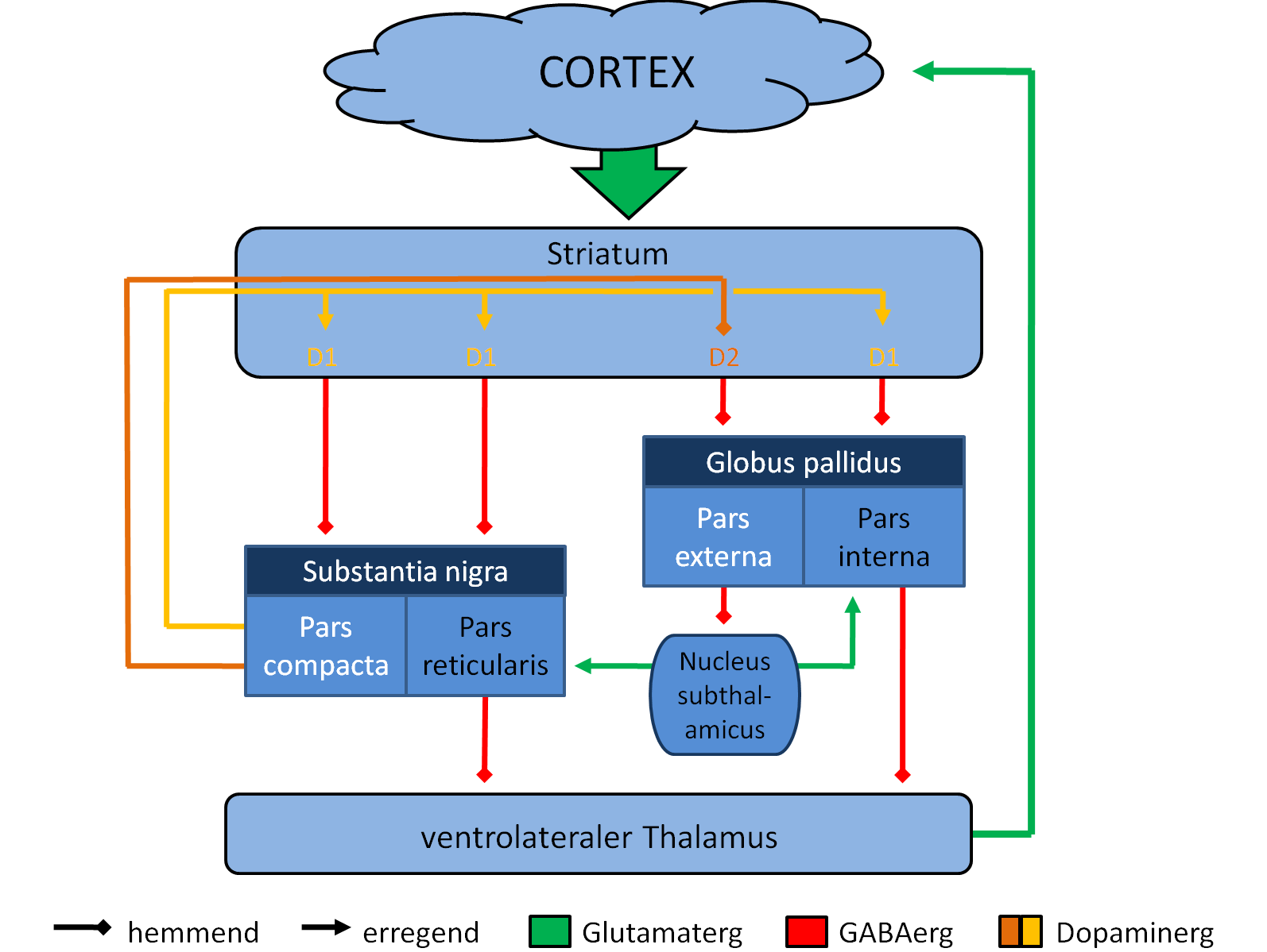
The main nucleus of the basal ganglia is the striatum which projects directly to the globus pallidus via a pathway of striatopallidal fibers. The striato-pallidal pathway has a whitish appearance due to the myelinated fibers. This projection comprises successively the external globus pallidus (GPe), the internal globus pallidus (GPi), the pars compacta of the substantia nigra (SNc), and the pars reticulata of substantia nigra (SNr). The neurons of this projection are inhibited by GABAergic synapses from the dorsal striatum. Among these targets, the GPe does not send axons outside the system. Others send axons to the superior colliculus. Two others comprise the output to the thalamus, forming two separate channels: one through the internal segment of the globus pallidus to the ventral oralis nuclei of the thalamus and from there to the cortical supplementary motor area and another through the substantia nigra to the ventral anterior nuclei of the thalamus and from there to the frontal cortex and the occulomotor cortex.
Blood supply
Deep penetrating striate arteries supply blood to the striatum. These arteries include the recurrent artery of Heubner arising from the anterior cerebral artery, and the lenticulostriate arteries arising from the middle cerebral artery.
Function
The ventral striatum, and the nucleus accumbens in particular, primarily mediates reward, cognition, reinforcement, and motivational salience. By contrast, the dorsal striatum primarily mediates cognition involving motor function, certain executive functions (e.g., inhibitory control and impulsivity), and stimulus-response learning. There is a small degree of overlap, as the dorsal striatum is also a component of the reward system that, along with the nucleus accumbens core, mediates the encoding of new motor programs associated with future reward acquisition (e.g., the conditioned motor response to a reward cue).
The striatum is also thought to play a role in an at least partially dissociable executive control network for language, applied to both verbal working memory and verbal attention. These models take the form of a frontal-striatal network for language processing. While the striatum is often not included in models of language processing, as most models only include cortical regions, integrative models are becoming more popular in light of imaging studies, lesion studies on aphasic patients, and studies of language disorders concomitant with diseases known to affect the striatum like Parkinson's and Huntington's disease.
Metabotropic dopamine receptors are present both on spiny neurons and on cortical axon terminals. Second messenger cascades triggered by activation of these dopamine receptors can modulate pre- and postsynaptic function, both in the short term and in the long term. In humans, the striatum is activated by stimuli associated with reward, but also by aversive, novel, unexpected, or intense stimuli, and cues associated with such events. fMRI evidence suggests that the common property linking these stimuli, to which the striatum is reacting, is salience under the conditions of presentation. A number of other brain areas and circuits are also related to reward, such as frontal areas. Functional maps of the striatum reveal interactions with widely distributed regions of the cerebral cortex important to a diverse range of functions.
The interplay between the striatum and the prefrontal cortex is relevant for behavior, particularly adolescent development as proposed by the dual systems model.
Clinical significance
Parkinson's disease and other movement disorders
Parkinson's disease results in loss of dopaminergic innervation to the dorsal striatum (and other basal ganglia) and a cascade of consequences. Atrophy of the striatum is also involved in Huntington's disease, and movement disorders such as chorea, choreoathetosis, and dyskinesias. These have also been described as circuit disorders of the basal ganglia.
Addiction

Addiction, a disorder of the brain's reward system, arises through the overexpression of DeltaFosB (ΔFosB), a transcription factor, in the D1-type medium spiny neurons of the ventral striatum. ΔFosB is an inducible gene which is increasingly expressed in the nucleus accumbens as a result of repeatedly using an addictive drug or overexposure to other addictive stimuli.
Schizophrenia spectrum disorders
The mesolimbic hypothesis of schizophrenia has long emphasized the role of hyperdopaminergia in the mesolimbic pathway, which extends from the ventral tegmental area to the ventral striatum. Abnormally elevated dopaminergic transmission in this pathway has been associated with the emergence of positive symptoms, such as hallucinations and delusions. Most antipsychotic treatments exert their effects by reducing dopamine binding to receptors in this region. More recent evidence, however, suggests that the pathway from the substantia nigra to the dorsal striatum may also play a significant role in the pathophysiology of schizophrenia. This has led to the development of the mesostriatal hypothesis, which expands the focus beyond the ventral striatum to include dopaminergic dysfunction in the dorsal components of the striatum and could help accounting for the negative and cognitive symptoms.
Bipolar disorder
An association has been observed between striatal expression of variants of the PDE10A gene and some bipolar I disorder patients. Variants of other genes, DISC1 and GNAS, have been associated with bipolar II disorder.
Autism spectrum disorder
Autism spectrum disorder (ASD) is characterized by cognitive inflexibility and poor understanding of social systems. This inflexible behavior originates in defects in the prefrontal cortex as well as the striatal circuits. The defects in the striatum seem to specifically contribute to the motor, social and communication impairments seen in ASD patients. In mice which have an ASD-like phenotype induced via the overexpression of the eukaryotic initiation of translation factor 4E, it has been shown that these defects seem to stem from the reduced ability to store and process information in the striatum, which leads to the difficulty seen in forming new motor patterns, as well as disengaging from existing ones.
Dysfunction
Dysfunction in the ventral striatum can lead to a variety of disorders, most notably depression and obsessive-compulsive disorder. Because of its involvement in reward pathways, the ventral striatum has also been implicated in playing a critical role in addiction. It has been well established that the ventral striatum is strongly involved in mediating the reinforcing effects of drugs, especially stimulants, through dopaminergic stimulation.
Language disorders
Lesions to the striatum have been associated with deficits in speech production and comprehension. While striatal damage can impact all levels of language, damage can broadly be characterized as affecting the ability to manipulate linguistic units and rules, resulting in the promotion of default linguistic forms in conflicting situations in which selection, inhibition, and monitoring load is increased. Two subregions of the striatum have been shown to be particularly important in language: the caudate nucleus and left putamen. Lesions localized to the caudate nucleus, as well as direct electrical stimulation, can result in lexical paraphasias and perservations (continuations of an utterance after the stimulus has ceased), which is associated with inhibited executive control, in the sense that executive control allows for the selection of the best choice among competing alternatives. Stimulation of the putamen results in the inhibition of articulatory sequences and the inability to initiate motor speech commands.
History
In the seventeenth and eighteenth centuries, the term corpus striatum was used to designate many distinct, deep, infracortical elements of the hemisphere. Etymologically, it is derived from (Latin) striatus = "grooved, striated" and the English striated = having parallel lines or grooves on the surface. In 1876 David Ferrier contributed decades of research to the subject; concluding that the corpus striatum was vital in the "organization and generation of voluntary movement". In 1941, Cécile and Oskar Vogt simplified the nomenclature by proposing the term striatum for all elements in the basal ganglia built with striatal elements: the caudate nucleus, the putamen, and the fundus striati, which is the ventral part linking the two preceding together ventrally to the inferior part of the internal capsule.
The term neostriatum was coined by comparative anatomists comparing the subcortical structures between vertebrates, because it was thought to be a phylogenetically newer section of the corpus striatum. The term is still used by some sources, including Medical Subject Headings.
Other animals
In birds the term used was the paleostriatum augmentatum, while in the new avian terminology listing (as of 2002) for neostriatum this has been changed to the nidopallium.
In non-primate species, the islands of Calleja are included in the ventral striatum.
Additional images
File:Striatum coronal sections.gif|Striatum highlighted in green on coronal T1 MRI images File:Striatum sagittal sections.gif|Striatum highlighted in green on sagittal T1 MRI images File:Striatum transversal sections.gif|Striatum highlighted in green on transversal T1 MRI images
References
References
- "Basal ganglia".
- (February 2013). "The neurocircuitry of illicit psychostimulant addiction: acute and chronic effects in humans". Subst. Abuse Rehabil..
- "striatum {{!}} Definition of striatum in English by Oxford Dictionaries".
- (October 2019). "The Striatum's Role in Executing Rational and Irrational Economic Behaviors". Neuroscientist.
- (February 2014). "MR Anatomy of Deep Brain Nuclei with Special Reference to Specific Diseases and Deep Brain Stimulation Localization". The Neuroradiology Journal.
- "Striatum definition and meaning {{!}} Collins English Dictionary".
- "Ventral striatum – NeuroLex".
- "Ventral Striatum Definition – Medical Dictionary".
- "Ventral Striatum – Medical Definition".
- (9 March 2022). "Opposing Roles of the Dorsolateral and Dorsomedial Striatum in the Acquisition of Skilled Action Sequencing in Rats". The Journal of Neuroscience.
- (June 2019). "Role of basal ganglia neurocircuitry in the pathology of psychiatric disorders". Psychiatry and Clinical Neurosciences.
- (2013). "Fundamental neuroscience". Elsevier Academic Press.
- (2017). "The Striosome and Matrix Compartments of the Striatum: A Path through the Labyrinth from Neurochemistry toward Function". ACS Chemical Neuroscience.
- (December 2011). "Spontaneous firing and evoked pauses in the tonically active cholinergic interneurons of the striatum". Neuroscience.
- (July 2004). "Coincident but Distinct Messages of Midbrain Dopamine and Striatal Tonically Active Neurons". Neuron.
- (1 December 1995). "Regional, cellular, and subcellular variations in the distribution of D1 and D5 dopamine receptors in primate brain". The Journal of Neuroscience.
- (1996). "Neuronal synchronization of tonically active neurons in the striatum of normal and parkinsonian primates". Journal of Neurophysiology.
- (2020). "Polysynaptic inhibition between striatal cholinergic interneurons shapes their network activity patterns in a dopamine-dependent manner". Nature Communications.
- (May 1999). "Inhibitory control of neostriatal projection neurons by GABAergic interneurons". Nature Neuroscience.
- (19 May 2010). "Electrophysiological and Morphological Characteristics and Synaptic Connectivity of Tyrosine Hydroxylase-Expressing Neurons in Adult Mouse Striatum". Journal of Neuroscience.
- (16 November 2011). "A Novel Functionally Distinct Subtype of Striatal Neuropeptide Y Interneuron". Journal of Neuroscience.
- (11 December 2011). "GABAergic circuits mediate the reinforcement-related signals of striatal cholinergic interneurons". Nature Neuroscience.
- (2010). "Heterogeneity and Diversity of Striatal GABAergic Interneurons". Frontiers in Neuroanatomy.
- (February 2014). "Neurogenesis in the Striatum of the Adult Human Brain". Cell.
- (16 February 2016). "Adult neurogenesis in the human striatum: possible implications for psychiatric disorders". Molecular Psychiatry.
- (February 2010). "Forebrain neurogenesis after focal Ischemic and traumatic brain injury.". Neurobiology of Disease.
- (14 June 2006). "Subventricular Zone-Derived Neuroblasts Migrate and Differentiate into Mature Neurons in the Post-Stroke Adult Striatum". Journal of Neuroscience.
- (September 1999). "Anatomical re-evaluation of the corticostriatal projections to the caudate nucleus: a retrograde labeling study in the cat". Neuroscience Research.
- (2010). "Conditional Routing of Information to the Cortex: A Model of the Basal Ganglia's Role in Cognitive Coordination". Psychological Review.
- "Ventral striatum – NeuroLex".
- "Icahn School of Medicine {{!}} Neuroscience Department {{!}} Nestler Lab {{!}} Brain Reward Pathways".
- (April 1992). "Functions of dopamine in the dorsal and ventral striatum". Seminars in Neuroscience.
- (August 2015). "The ins and outs of the striatum: Role in drug addiction". Neuroscience.
- (June 2010). "Adenosine-cannabinoid receptor interactions. Implications for striatal function". Br. J. Pharmacol..
- (2016). "''In vivo'' Exploration of the Connectivity between the Subthalamic Nucleus and the Globus Pallidus in the Human Brain Using Multi-Fiber Tractography". Frontiers in Neuroanatomy.
- (2012). "Neuroscience".
- (2009). "Molecular Neuropharmacology: A Foundation for Clinical Neuroscience". McGraw-Hill Medical.
- (August 2021). "Striatum and language processing: Where do we stand?". Cognition.
- (March 1997). "A Neural Dissociation within Language: Evidence that the Mental Dictionary Is Part of Declarative Memory, and that Grammatical Rules Are Processed by the Procedural System". Journal of Cognitive Neuroscience.
- (2001). "The neurobiology of slow synaptic transmission". Science.
- (2014). "Local control of striatal dopamine release". Frontiers in Behavioral Neuroscience.
- UCL. (25 June 2008). "Adventure - it's all in the mind, say UCL neuroscientists".
- (2013). "New insights into the specificity and plasticity of reward and aversion encoding in the mesolimbic system". Journal of Neuroscience.
- (June 2004). "The Emergence of Collaborative Brain Function: fMRI Studies of the Development of Response Inhibition". Annals of the New York Academy of Sciences.
- "Department of Physiology, Development and Neuroscience: About the Department".
- (2012). "The organization of the human striatum estimated by intrinsic functional connectivity". Journal of Neurophysiology.
- (April 2010). "A dual systems model of adolescent risk-taking". Developmental Psychobiology.
- Walker FO. (January 2007). "Huntington's disease". [[The Lancet.
- (January 2007). "Circuits and Circuit Disorders of the Basal Ganglia". Archives of Neurology.
- Nestler EJ. (December 2013). "Cellular basis of memory for addiction". Dialogues Clin. Neurosci..
- (December 2011). "Natural rewards, neuroplasticity, and non-drug addictions". Neuropharmacology.
- (11 March 2010). "A Possible Role for the Striatum in the Pathogenesis of the Cognitive Symptoms of Schizophrenia". Neuron.
- (3 June 2020). "Subcortical Dopamine and Cognition in Schizophrenia: Looking Beyond Psychosis in Preclinical Models". Frontiers in Neuroscience.
- (1 August 2020). "Antipsychotics: Mechanisms underlying clinical response and side-effects and novel treatment approaches based on pathophysiology". Neuropharmacology.
- (17 October 2018). "Defining the Locus of Dopaminergic Dysfunction in Schizophrenia: A Meta-analysis and Test of the Mesolimbic Hypothesis". Schizophrenia Bulletin.
- (2 May 2025). "Functional connectivity of the striatum in psychosis: Meta-analysis of functional magnetic resonance imaging studies and replication on an independent sample". Neuroscience & Biobehavioral Reviews.
- (2 October 2012). "Genetic association of cyclic AMP signaling genes with bipolar disorder". Translational Psychiatry.
- (25 November 2009). "Probing Compulsive and Impulsive Behaviors, from Animal Models to Endophenotypes: A Narrative Review". Neuropsychopharmacology.
- (23 December 2012). "Exaggerated translation causes synaptic and behavioural aberrations associated with autism". Nature.
- (November 2013). "From the ventral to the dorsal striatum: Devolving views of their roles in drug addiction". Neuroscience & Biobehavioral Reviews.
- Fedorenko, Evelina. (2014). "The role of domain-general cognitive control in language comprehension". Frontiers in Psychology.
- (14 March 2000). "The anatomy of aphasia revisited". Neurology.
- (July 2005). "The role of dominant striatum in language: a study using intraoperative electrical stimulations". Journal of Neurology, Neurosurgery & Psychiatry.
- (2006-03-01). "Neural modeling and imaging of the cortical interactions underlying syllable production". Brain and Language.
- [[Raymond Vieussens]], 1685
- (16 August 2019). "Striatus".
- (9 November 2019). "Striated".
- (1998). "Neural systems for control of voluntary action ? A hypothesis". Advances in Biophysics.
- (23 September 1994). "The Basal Ganglia and Adaptive Motor Control". Science.
- (December 2010). "Dopamine in Motivational Control: Rewarding, Aversive, and Alerting". Neuron.
- Ferrier, David. (1877-07-01). "Ferrier on the Functions of the Brain". The British and Foreign Medico-Chirurgical Review.
- (June 2012). "Striatal Mechanisms Underlying Movement, Reinforcement, and Punishment". Physiology.
- "NeuroNames Ancillary: fundus striati".
- {{MeshName. Neostriatum
- "New Terminology for the Neostriatum".
This article was imported from Wikipedia and is available under the Creative Commons Attribution-ShareAlike 4.0 License. Content has been adapted to SurfDoc format. Original contributors can be found on the article history page.
Ask Mako anything about Striatum — get instant answers, deeper analysis, and related topics.
Research with MakoFree with your Surf account
Create a free account to save articles, ask Mako questions, and organize your research.
Sign up freeThis content may have been generated or modified by AI. CloudSurf Software LLC is not responsible for the accuracy, completeness, or reliability of AI-generated content. Always verify important information from primary sources.
Report