From Surf Wiki (app.surf) — the open knowledge base
Poliovirus
Enterovirus

Enterovirus
- PV-2 (Poliovirus-2)
- PV-3 (Poliovirus-3) Poliovirus, the causative agent of polio (also known as poliomyelitis), is a serotype of the species Enterovirus C, in the family of Picornaviridae. There are three poliovirus serotypes, numbered 1, 2, and 3.
Poliovirus is composed of an RNA genome and a protein capsid. The genome is a single-stranded positive-sense RNA (+ssRNA) genome that is about 7500 nucleotides long. The viral particle is about 30 nm in diameter with icosahedral symmetry. Because of its short genome and its simple composition—only a strand of RNA and a nonenveloped icosahedral protein coat encapsulating it—poliovirus is widely regarded as the simplest significant virus.
Poliovirus is one of the most well-characterized viruses, and has become a useful model system for understanding the biology of RNA viruses.
Replication cycle
Poliovirus infects human cells by binding to an immunoglobulin-like receptor, CD155 (also known as the poliovirus receptor or PVR) on the cell surface. Interaction of poliovirus and CD155 facilitates an irreversible conformational change of the viral particle necessary for viral entry. Following attachment to the host cell membrane, entry of the viral nucleic acid was thought to occur one of two ways: via the formation of a pore in the plasma membrane through which the RNA is then "injected" into the host cell cytoplasm, or via virus uptake by receptor-mediated endocytosis. Recent experimental evidence supports the latter hypothesis and suggests that poliovirus binds to CD155 and is taken up by endocytosis. Immediately after internalization of the particle, the viral RNA is released.
Poliovirus is a positive-stranded RNA virus. Thus, the genome enclosed within the viral particle can be used as messenger RNA and immediately translated by the host cell. On entry, the virus hijacks the cell's translation machinery, causing inhibition of cellular protein synthesis in favor of virus-specific protein production. Unlike the host cell's mRNAs, the 5' end of poliovirus RNA is extremely long—over 700 nucleotides—and highly structured. This region of the viral genome is called an internal ribosome entry site (IRES). This region consists of many secondary structures and 3 or 4 domains. Domain 3 is a self folding RNA element that contains conserved structural motifs in various stable stem loops linked by two four-way junctions. As IRES consists of many domains, these domains themselves consist of many loops that contribute to modified translation without a 5’ end cap by hijacking ribosomes. The interaction loop of domain 3 is known as GNRA tetraloop. The residues of adenosines A180 and A181 in the GUAA tetraloop form hydrogen bonds via non canonical base pairing interactions with the base pairs of the receptors C230/G242 and G231/C241, respectively. Genetic mutations in this region prevent viral protein production. The first IRES to be discovered was found in poliovirus RNA.
Poliovirus mRNA is translated as one long polypeptide. This polypeptide is then autocleaved by internal proteases into about 10 individual viral proteins. Not all cleavages occur with the same efficiency. Therefore, the amounts of proteins produced by the polypeptide cleavage vary: for example, smaller amounts of 3Dpol are produced than those of capsid proteins, VP1–4. These individual viral proteins are:

- 3Dpol, an RNA dependent RNA polymerase whose function is to make multiple copies of the viral RNA genome
- 2Apro and 3Cpro/3CDpro, proteases which cleave the viral polypeptide
- VPg (3B), a small protein that binds viral RNA and is necessary for synthesis of viral positive and negative strand RNA
- 2BC, 2B, 2C (an ATPase), 3AB, 3A, 3B proteins which comprise the protein complex needed for virus replication.
- VP0, which is further cleaved into VP2 and VP4, VP1 and VP3, proteins of the viral capsid
After translation, transcription and genome replication which involve a single process, synthesis of (+) RNA) is realized. For the infecting (+)RNA to be replicated, multiple copies of (−)RNA must be transcribed and then used as templates for (+)RNA synthesis. Replicative intermediates (RIs), which are an association of RNA molecules consisting of a template RNA and several growing RNAs of varying length, are seen in both the replication complexes for (−)RNAs and (+)RNAs. For synthesis of each negative-strand and positive-strand RNAs, VPg protein in the poliovirus works as a primer. RNA-dependent RNA polymerase of the poliovirus adds two uracil nucleotides (UU) to VPg protein utilizing the poly(A) tail at the 3′-end of the +ssRNA genome as a pattern for synthesis of the negative-strand antigenomic RNA. To initiate this −ssRNA synthesis, the tyrosine hydroxyl of VPg is needed. But for the initiation of positive strand RNA synthesis, CRE-dependent VPg uridylylation is needed. Which means that VPg is once more utilized as a primer however this time it adds the two uridine triphosphates using a cis-acting replication element (CRE) as a template.
The CRE of poliovirus is identified as an unachieved base-paired stem and a final loop consisting of 61 nt. The CRE is found in enteroviruses. It is a highly preserved secondary RNA structural element and bedded in the genome's polyprotein-coding region. The complex can be translocated to the 5' region of the genome that have no coding activity, at least 3.7-kb distant from the initial location. This process can occurs without negatively influencing activity. CRE copies do not influence replication negatively. Uridylylation process of VPg that takes place at CRE needs the presence of 3CDpro that is an RNA binding protein. It is attached to the CRE directly and specifically. Because of its presence VPg can bind the CRE properly and primary production proceeds without problems.
Some of the (+) RNA molecules are used as templates for further (−) RNA synthesis, some function as mRNA, and some are destined to be the genomes of progeny virions.
In the assembly of new virus particles (i.e. the packaging of progeny genome into a procapsid which can survive outside the host cell), including, respectively:
- Five copies each of VP0, VP3, and VP1 whose N termini and VP4 form interior surface of capsid, assemble into a 'pentamer' and 12 pentamers form a procapsid. (The outer surface of capsid is consisting of VP1, VP2, VP3; C termini of VP1 and VP3 form the canyons which around each of the vertices; around this time, the 60 copies of VP0 are cleaved into VP4 and VP2.)
- Each procapsid acquires a copy of the virus genome, with VPg still attached at the 5' end.
Fully assembled poliovirus leaves the confines of its host cell by lysis 4 to 6 hours following initiation of infection in cultured mammalian cells. The mechanism of viral release from the cell is unclear, but each dying cell can release up to 10,000 polio virions.
Drake demonstrated that poliovirus is able to undergo multiplicity reactivation. That is, when polioviruses were irradiated with UV light and allowed to undergo multiple infections of host cells, viable progeny could be formed even at UV doses that inactivated the virus in single infections. Poliovirus can undergo genetic recombination when at least two viral genomes are present in the same host cell. Kirkegaard and Baltimore presented evidence that RNA-dependent RNA polymerase (RdRP) catalyzes recombination by a copy choice mechanism in which the RdRP switches between (+)ssRNA templates during negative strand synthesis. Recombination in RNA viruses appears to be an adaptive mechanism for repairing genome damage.
Origin and serotypes
Poliovirus is structurally similar to other human enteroviruses (coxsackieviruses, echoviruses, and rhinoviruses), which also use immunoglobulin-like molecules to recognize and enter host cells. The distinct speciation of poliovirus probably occurred as a result of a change in cellular receptor specificity from intercellular adhesion molecule-1 (ICAM-1) (used by C-cluster Coxsackie A viruses) to CD155, leading to a change in pathogenicity and allowing the virus to infect nerve tissue.
The mutation rate in the virus is relatively high even for an RNA virus, with a synonymous substitution rate of 1.0 × 10⁻² substitutions/site/year and a non-synonymous substitution rate of 3.0 × 10⁻⁴ substitutions/site/year. Base distribution within the genome is not random; adenosine is less common than expected at the 5' end and higher at the 3' end. Codon use is not random; codons ending in adenosine are favoured, and those ending in cytosine or guanine are avoided. Codon use differs between the three genotypes, and appears to be driven by mutation rather than selection.
The three serotypes of poliovirus, PV-1, PV-2, and PV-3, each have a slightly different capsid protein. Capsid proteins define cellular receptor specificity and virus antigenicity. PV-1 is the most common form encountered in nature, but all three forms are extremely infectious. and in October 2019 for wild PV-3 after last being detected in 2012. However, circulating vaccine-derived poliovirus (variant poliovirus, cVDPV) of all three serotypes continues to circulate and cause paralysis, having been detected in 32 countries in 2023.
Specific strains of each serotype are used to prepare vaccines against polio. Inactive polio vaccine is prepared by formalin inactivation of three wild, virulent reference strains: Mahoney or Brunenders (PV-1), MEF-1/Lansing (PV-2), and Saukett/Leon (PV-3). Oral polio vaccine contains live attenuated (weakened) strains of the three serotypes of poliovirus. Passaging the virus strains in monkey kidney epithelial cells introduces mutations in the viral IRES, and hinders (or attenuates) the ability of the virus to infect nerve tissue.
Polioviruses were formerly classified as a distinct species belonging to the genus Enterovirus in the family Picornaviridae. In 2008, Poliovirus ceased to be recognized as a species, and the three serotypes were assigned to the species Human enterovirus C (later renamed Enterovirus C) in the genus Enterovirus in the family Picornaviridae. The type species of the genus Enterovirus was changed from Poliovirus to (Human) Enterovirus C.
Pathogenesis
Main article: Polio
The primary determinant of infection for any virus is its ability to enter a cell and produce additional infectious particles. The presence of CD155 is thought to define the animals and tissues that can be infected by poliovirus. CD155 is found (outside of laboratories) only on the cells of humans, higher primates, and Old World monkeys. Poliovirus is, however, strictly a human pathogen, and does not naturally infect any other species (although chimpanzees and Old World monkeys can be experimentally infected).
The CD155 gene appears to have been subject to positive selection. The protein has several domains of which domain D1 contains the polio virus binding site. Within this domain, 37 amino acids are responsible for binding the virus.
Poliovirus is an enterovirus. Infection occurs via the fecal–oral route, meaning that one ingests the virus and viral replication occurs in the gastrointestinal tract. Virus is shed in the feces of infected individuals. In 95% of cases only a primary, transient presence of viremia (virus in the bloodstream) occurs, and the poliovirus infection is asymptomatic. In about 5% of cases, the virus spreads and replicates in other sites such as brown fat, reticuloendothelial tissue, and muscle. The sustained viral replication causes secondary viremia and leads to the development of minor symptoms such as fever, headache, and sore throat. Paralytic poliomyelitis occurs in less than 1% of poliovirus infections. Paralytic disease occurs when the virus enters the central nervous system (CNS) and replicates in motor neurons within the spinal cord, brain stem, or motor cortex, resulting in the selective destruction of motor neurons leading to temporary or permanent paralysis. This is a very rare event in babies, who still have anti-poliovirus antibodies acquired from their mothers. In rare cases, paralytic poliomyelitis leads to respiratory arrest and death. In cases of paralytic disease, muscle pain and spasms are frequently observed prior to onset of weakness and paralysis. Paralysis typically persists from days to weeks prior to recovery.
In many respects, the neurological phase of infection is thought to be an accidental diversion of the normal gastrointestinal infection. A second hypothesis suggests that the virions are transported from peripheral tissues that have been bathed in the viremic blood, for example muscle tissue, to the spinal cord through nerve pathways via retrograde axonal transport. A third hypothesis is that the virus is imported into the CNS via infected monocytes or macrophages.
Poliomyelitis is a disease of the central nervous system. However, CD155 is believed to be present on the surface of most or all human cells. Therefore, receptor expression does not explain why poliovirus preferentially infects certain tissues. This suggests that tissue tropism is determined after cellular infection. Recent work has suggested that the type I interferon response (specifically that of interferon alpha and beta) is an important factor that defines which types of cells support poliovirus replication. In mice expressing CD155 (through genetic engineering) but lacking the type I interferon receptor, poliovirus not only replicates in an expanded repertoire of tissue types, but these mice are also able to be infected orally with the virus.
Immune system avoidance
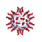
Reconstructed image from cryo-electron microscopy.]]
Poliovirus has two key mechanisms to evade the immune system. First, it can survive the highly acidic conditions of the stomach, allowing ingested viruses to infect the host and spread throughout the body via the lymphatic system. Second, because it can replicate very quickly, the virus overwhelms the host's organs before an immune response can be mounted. If detail is given at the attachment phase; poliovirus with canyons on the virion surface have virus attachment sites located in pockets at the canyon bases. The canyons are too narrow for access by antibodies, so the virus attachment sites are protected from the host's immune surveillance, while the remainder of the virion surface can mutate to avoid the host's immune response.
Individuals who are exposed to poliovirus, either through infection or by immunization with polio vaccine, develop immunity. In immune individuals, antibodies against poliovirus are present in the tonsils and gastrointestinal tract (specifically IgA antibodies) and are able to block poliovirus replication; IgG and IgM antibodies against poliovirus can prevent the spread of the virus to motor neurons of the central nervous system.
Eradication
Research
Transgenic mice
Although humans are the only known natural hosts of poliovirus, monkeys can be experimentally infected and they have long been used to study poliovirus. In 1990–91, a small animal model of poliomyelitis was developed by two laboratories. Mice were engineered to express a human receptor to poliovirus (hPVR).
Unlike normal mice, transgenic poliovirus receptor (TgPVR) mice are susceptible to poliovirus injected intravenously or intramuscularly, and when injected directly into the spinal cord or the brain. Upon infection, TgPVR mice show signs of paralysis that resemble those of poliomyelitis in humans and monkeys, and the central nervous systems of paralyzed mice are histocytochemically similar to those of humans and monkeys. This mouse model of human poliovirus infection has proven to be an invaluable tool in understanding poliovirus biology and pathogenicity.
Three distinct types of TgPVR mice have been well studied:
- In TgPVR1 mice, the transgene encoding the human PVR was incorporated into mouse chromosome 4. These mice express the highest levels of the transgene and the highest sensitivity to poliovirus. TgPVR1 mice are susceptible to poliovirus through the intraspinal, intracerebral, intramuscular, and intravenous pathways, but not through the oral route.
- TgPVR21 mice have incorporated the human PVR at chromosome 13. These mice are less susceptible to poliovirus infection through the intracerebral route, possibly because they express decreased levels of hPVR. TgPVR21 mice have been shown to be susceptible to poliovirus infection through intranasal inoculation, and may be useful as a mucosal infection model.
- In TgPVR5 mice, the human transgene is located on chromosome 12. These mice exhibit the lowest levels of hPVR expression and are the least susceptible to poliovirus infection.
- A fourth TgPVR mouse model - these "cPVR" mice carry hPVR cDNA, driven by a β-actin promoter, and have proven susceptible to poliovirus through intracerebral, intramuscular, and intranasal routes. In addition, these mice are capable of developing the bulbar form of polio after intranasal inoculation.
The development of the TgPVR mouse has had a profound effect on oral poliovirus vaccine (OPV) production. Previously, monitoring the safety of OPV had to be performed using monkeys, because only primates are susceptible to the virus. In 1999, the World Health Organization approved the use of the TgPVR mouse as an alternative method of assessing the effectiveness of the vaccine against poliovirus type-3. In 2000, the mouse model was approved for tests of vaccines against type-1 and type-2 poliovirus.
Genetic modification
A modification of the poliovirus, called PVSRIPO, was tested in early clinical trials as a possible treatment for cancer. , a number of clinical trials are underway.
A drawback of the attenuated virus used in the Sabin oral polio vaccine is its potential to cause vaccine-associated paralytic poliomyelitis (VAPP) in approximately one individual per every 2.7million doses administered. In addition, the live virus can also circulate in under-vaccinated populations (circulating vaccine-derived poliovirus, cVDPV) and over time can revert to a neurovirulent form causing paralytic polio. Researchers have developed a poliovirus type 2 vaccine strain that is genetically more stable and less likely to regain virulence than the original Sabin2 strain, with three key genetic modifications. The vaccine derived from this strain, novel oral polio virus type 2 (nOPV2), was granted emergency licencing in 2021, and subsequently full licensure in December 2023. Genetically stabilsed vaccines targeting poliovirus types 1 and 3 are in development, with the intention that these will eventually completely replace the Sabin vaccines.
History
Discovery
Poliovirus was first isolated in 1909 by Karl Landsteiner and Erwin Popper. The structure of the virus was first elucidated in 1958 using X-ray diffraction by a team at Birkbeck College led by Rosalind Franklin, showing the polio virus to have icosahedral symmetry.
Genome
In 1981, the poliovirus genome was published by two different teams of researchers: by Vincent Racaniello and David Baltimore at MIT and by Naomi Kitamura and Eckard Wimmer at Stony Brook University.
Structure
The three-dimensional structure of poliovirus was determined in 1985 by James Hogle at Scripps Research Institute using X-ray crystallography.
Cloning

In 1981, Racaniello and Baltimore used recombinant DNA technology to generate the first infectious clone of an animal RNA virus, poliovirus. DNA encoding the RNA genome of poliovirus was introduced into cultured mammalian cells and infectious poliovirus was produced. Creation of the infectious clone propelled understanding of poliovirus biology, and has become a standard technology used to study many other viruses.
Synthesis
In 2002, Eckard Wimmer's group at Stony Brook University succeeded in synthesizing poliovirus from its chemical code, producing the world's first synthetic virus. Scientists first converted poliovirus's published RNA sequence, 7741 bases long, into a DNA sequence, as DNA was easier to synthesize. Short fragments of this DNA sequence were obtained by mail-order, and assembled. The complete viral genome was then assembled by a gene synthesis company. Nineteen markers were incorporated into the synthesized DNA, so that it could be distinguished from natural poliovirus. Enzymes were used to convert the DNA back into RNA, its natural state. Other enzymes were then used to translate the RNA into a polypeptide, producing functional viral particle. This whole painstaking process took two years. The newly minted synthetic virus was injected into PVR transgenic mice, to determine if the synthetic version was able to cause disease. The synthetic virus was able to replicate, infect, and cause paralysis or death in mice. However, the synthetic version was between 1,000 and 10,000 times weaker than the original virus, probably due to one of the added markers.
References
References
- (August 2021). "Sherris Medical Microbiology". McGraw Hill.
- (2002). "Poliovirus cell entry: common structural themes in viral cell entry pathways". Annual Review of Microbiology.
- (August 2021). "The machinery of life". Copernicus.
- (July 2007). "Epidemics to eradication: the modern history of poliomyelitis". Virology Journal.
- (March 1989). "Cellular receptor for poliovirus: molecular cloning, nucleotide sequence, and expression of a new member of the immunoglobulin superfamily". Cell.
- (April 2003). "Complexes of poliovirus serotypes with their common cellular receptor, CD155". Journal of Virology.
- (October 1969). "Entry and release of poliovirus as observed by electron microscopy of cultured cells". Journal of Virology.
- (October 1990). "Neutralization of poliovirus by cell receptors expressed in insect cells". Journal of Virology.
- (November 1993). "Characterization of poliovirus conformational alteration mediated by soluble cell receptors". Virology.
- (August 2005). "Poliovirus and poliomyelitis: a tale of guts, brains, and an accidental event". Virus Research.
- (July 2007). "Imaging poliovirus entry in live cells". PLOS Biology.
- (1962). "Virus specific protein and a ribo-nucleic acid associated with ribosomes in poliovirus infected HeLa cells". Cold Spring Harbor Symposia on Quantitative Biology.
- (February 2016). "Modeling Three-Dimensional Structural Motifs of Viral IRES". Journal of Molecular Biology.
- (April 1995). "Initiation of protein synthesis by the eukaryotic translational apparatus on circular RNAs". Science.
- (July 1988). "Internal initiation of translation of eukaryotic mRNA directed by a sequence derived from poliovirus RNA". Nature.
- (August 1988). "A segment of the 5' nontranslated region of encephalomyocarditis virus RNA directs internal entry of ribosomes during in vitro translation". Journal of Virology.
- (2007). "Virology: Principles and Applications". John Wiley & Sons.
- (August 2021). "Viruses: Biology, Applications, and Control". Garland Science.
- "Poliomyelitis". Brown University.
- (2007). "Virology: Principles and Applications". John Wiley & Sons.
- (2016). "Essential Human Virology". Elsevier.
- (April 2003). "Poliovirus CRE-dependent VPg uridylylation is required for positive-strand RNA synthesis but not for negative-strand RNA synthesis". Journal of Virology.
- (January 2003). "Structure and function analysis of the poliovirus cis-acting replication element (CRE)". RNA.
- (2007). "Virology: Principles and Applications". John Wiley & Sons.
- (2007). "Virology: Principles and Applications". John Wiley & Sons.
- (2005). "Vaccine-derived polioviruses and the endgame strategy for global polio eradication". Annual Review of Microbiology.
- (August 1958). "Interference and multiplicity reactivation in polioviruses". Virology.
- (November 1986). "The mechanism of RNA recombination in poliovirus". Cell.
- (June 2010). "How RNA viruses maintain their genome integrity". The Journal of General Virology.
- (January 2018). "Sex in microbial pathogens". Infection, Genetics and Evolution.
- (May 2007). "Evidence for emergence of diverse polioviruses from C-cluster coxsackie A viruses and implications for global poliovirus eradication". Proceedings of the National Academy of Sciences of the United States of America.
- (May 2008). "Calibration of multiple poliovirus molecular clocks covering an extended evolutionary range". Journal of Virology.
- (December 1981). "Mononucleotide and dinucleotide frequencies, and codon usage in poliovirion RNA". Nucleic Acids Research.
- (March 2011). "Analysis of codon usage and nucleotide composition bias in polioviruses". Virology Journal.
- (2015-09-20). "Global eradication of wild poliovirus type 2 declared". [[Global Polio Eradication Initiative]].
- "GPEI-Two out of three wild poliovirus strains eradicated".
- (May 2024). "Progress Toward Poliomyelitis Eradication - Worldwide, January 2022-December 2023". MMWR. Morbidity and Mortality Weekly Report.
- (July 2009). "Ratification vote on taxonomic proposals to the International Committee on Taxonomy of Viruses (2008)". Archives of Virology.
- (August 2003). "Recruitment of nectin-3 to cell-cell junctions through trans-heterophilic interaction with CD155, a vitronectin and poliovirus receptor that localizes to alpha(v)beta3 integrin-containing membrane microdomains". The Journal of Biological Chemistry.
- (May 2006). "Ancient positive selection on CD155 as a possible cause for susceptibility to poliovirus infection in simians". Gene.
- (1969). "Polioviruse". Lippincott.
- (June 1956). "Pathogenesis of poliomyelitis; reappraisal in the light of new data". Science.
- (2007). "Virology: Principles and Applications". John Wiley & Sons.
- {{EMedicine. article. 306440. Acute Poliomyelitis
{{EMedicine. article. 967950. Pediatric Poliomyelitis - (March 1997). "Efficient delivery of circulating poliovirus to the central nervous system independently of poliovirus receptor". Virology.
- (October 1998). "Retrograde transport of intact poliovirus through the axon via the fast transport system". Virology.
- (October 1992). "Poliovirus spreads from muscle to the central nervous system by neural pathways". The Journal of Infectious Diseases.
- (March 2010). "Limited trafficking of a neurotropic virus through inefficient retrograde axonal transport and the type I interferon response". PLOS Pathogens.
- (April 2005). "The alpha/beta interferon response controls tissue tropism and pathogenicity of poliovirus". Journal of Virology.
- (August 2007). "Establishment of a poliovirus oral infection system in human poliovirus receptor-expressing transgenic mice that are deficient in alpha/beta interferon receptor". Journal of Virology.
- (January 2006). "One hundred years of poliovirus pathogenesis". Virology.
- (2007). "Virology: Principles and Applications". John Wiley & Sons.
- "WHO | Poliomyelitis".
- (October 1990). "Transgenic mice expressing a human poliovirus receptor: a new model for poliomyelitis". Cell.
- (February 1991). "Transgenic mice susceptible to poliovirus". Proceedings of the National Academy of Sciences of the United States of America.
- (February 1994). "Transgenic mice carrying the human poliovirus receptor: new animal models for study of poliovirus neurovirulence". Journal of Virology.
- (October 2001). "Recent insights into poliovirus pathogenesis". Trends in Microbiology.
- (1994). "Characterization of three different transgenic mouse lines that carry human poliovirus receptor gene--influence of the transgene expression on pathogenesis". Archives of Virology.
- (March 2004). "A poliomyelitis model through mucosal infection in transgenic mice bearing human poliovirus receptor, TgPVR21". Virology.
- (2003). "Transgenic mice as an alternative to monkeys for neurovirulence testing of live oral poliovirus vaccine: validation by a WHO collaborative study". Bulletin of the World Health Organization.
- (November 2014). "Oncolytic polio virotherapy of cancer". Cancer.
- (2022-09-01). "Update on Recombinant Poliovirus Cancer Immunotherapy".
- UCSF. (2017-06-01). "UCSF Glioma Trial: PVSRIPO in Recurrent Malignant Glioma".
- "OPV - Oral Polio Vaccine".
- (May 2020). "Engineering the Live-Attenuated Polio Vaccine to Prevent Reversion to Virulence". Cell Host & Microbe.
- (12 April 2024). "GPEI-OPV Oral polio vaccine".
- (2023-06-14). "Two New Vaccines Join the Fight to Eradicate Polio".
- (August 2021). "A History of Poliomyelitis". Yale University Press.
- (2019-03-14). "Behind the picture: Rosalind Franklin and the polio model". Medical Research Council.
- (2003). "Rosalind Franklin: The Dark Lady of DNA". Harper Collins.
- (2007). "J.D. Bernal: The Sage of Science". Oxford University Press.
- (August 1981). "Molecular cloning of poliovirus cDNA and determination of the complete nucleotide sequence of the viral genome". Proceedings of the National Academy of Sciences of the United States of America.
- (June 1981). "Primary structure, gene organization and polypeptide expression of poliovirus RNA". Nature.
- (September 1985). "Three-dimensional structure of poliovirus at 2.9 A resolution". Science.
- (November 1981). "Cloned poliovirus complementary DNA is infectious in mammalian cells". Science.
- (August 2002). "Chemical synthesis of poliovirus cDNA: generation of infectious virus in the absence of natural template". Science.
- (July 2002). "Virology. Active poliovirus baked from scratch". Science.
This article was imported from Wikipedia and is available under the Creative Commons Attribution-ShareAlike 4.0 License. Content has been adapted to SurfDoc format. Original contributors can be found on the article history page.
Ask Mako anything about Poliovirus — get instant answers, deeper analysis, and related topics.
Research with MakoFree with your Surf account
Create a free account to save articles, ask Mako questions, and organize your research.
Sign up freeThis content may have been generated or modified by AI. CloudSurf Software LLC is not responsible for the accuracy, completeness, or reliability of AI-generated content. Always verify important information from primary sources.
Report