From Surf Wiki (app.surf) — the open knowledge base
Oncocytoma
Tumor composed of oncocytes (epithelial cells with excess mitochondria)
Tumor composed of oncocytes (epithelial cells with excess mitochondria)
| Field | Value |
|---|---|
| name | Oncocytoma |
| caption | Micrograph of a parotid gland oncocytoma (right of image). Normal parotid gland is also present (left of image). H&E stain. |
An oncocytoma is a tumor made up of oncocytes, epithelial cells characterized by an excessive amount of mitochondria, resulting in an abundant acidophilic, granular cytoplasm. The cells and the tumor that they compose are often benign but sometimes may be premalignant or malignant.
Presentation
An oncocytoma is an epithelial tumor composed of oncocytes, large eosinophilic cells having small, round, benign-appearing nuclei with large nucleoli.
Oncocytoma can arise in a number of organs.
Renal oncocytoma
Main article: Renal oncocytoma
Renal oncocytoma is thought to arise from the intercalated cells of collecting ducts of the kidney. It represents 5% to 15% of surgically resected renal neoplasms.
Salivary gland oncocytoma
An salivary gland oncocytoma (also known as an oxyphilic adenoma) is a well-circumscribed, benign neoplastic growth comprising about one percent of all salivary gland tumors. The histopathology is marked by sheets of large, swollen polyhedral epithelial oncocytes, which are granular acidophilic parotid cells with centrally located nuclei. The granules are created by the mitochondria.
Symptoms
Salivary gland oncocytomas, 85 to 90 percent of which are located in the parotid gland, are firm, slowly growing, painless masses of less than 4 cm and may be bilateral. They are most common in females age 70 to 80.
Thyroid oncocytoma
Thyroid oncocytomas (also known as Hürthle cell tumours) can be benign (adenomas) or malignant (carcinomas). Grossly, oncocytic adenomas are encapsulated, solid nodules with a characteristic brown cut surface. The gross appearance of a minimally invasive oncocytic carcinoma is indistinguishable to that of an adenoma, while widely invasive oncocytic carcinomas are obviously invasive macroscopically and display pervasive vascular invasion with multifocal involvement of the thyroid gland. There are no reliable cytologic features which distinguish oncocytic adenomas from carcinomas and the only criterion for a diagnosis of malignancy is the identification of transcapsular or vascular invasion.
Symptoms
Patients with thyroid oncocytomas present with a thyroid nodule, usually with normal thyroid function. If the tumor is big or invasive, there may be other symptoms such as difficulty swallowing or talking.
Additional images
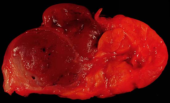
Image:Renal oncocytoma3.jpg|Micrograph of a renal oncocytoma. H&E stain. Image:Renal oncocytoma4.jpg|Micrograph of a renal oncocytoma. H&E stain. Image:Renal oncocytoma.jpg|Gross appearance of the cut surface of a nephrectomy specimen containing a renal oncocytoma. Note the rounded contour, the mahogany colour and the central scar.
References
References
- (April 2007). "Cutaneous oncocytoma - a report of three cases and review of the literature". Journal of Cutaneous Pathology.
- "Atlas of Genetics and Cytogenetics in Oncology and Haematology - Thyroid:oncocytic tumors".
This article was imported from Wikipedia and is available under the Creative Commons Attribution-ShareAlike 4.0 License. Content has been adapted to SurfDoc format. Original contributors can be found on the article history page.
Ask Mako anything about Oncocytoma — get instant answers, deeper analysis, and related topics.
Research with MakoFree with your Surf account
Create a free account to save articles, ask Mako questions, and organize your research.
Sign up freeThis content may have been generated or modified by AI. CloudSurf Software LLC is not responsible for the accuracy, completeness, or reliability of AI-generated content. Always verify important information from primary sources.
Report