From Surf Wiki (app.surf) — the open knowledge base
Iris dilator muscle
Smooth muscle of the eye
Smooth muscle of the eye
| Field | Value |
|---|---|
| Name | Iris dilator muscle |
| Latin | musculus dilatator pupillae |
| Image | Gray878.png |
| Caption | Iris, front view. (Muscle visible but not labeled.) |
| Image2 | Gray883.png |
| Caption2 | The upper half of a sagittal section through the front of the eyeball. (Iris dilator muscle is NOT labeled and not to be confused with "Radiating fibers" labeled near center, which are part of the ciliary muscle.) |
| Origin | Outer margins of iris |
| Insertion | Inner margins of iris |
| Nerve | Long ciliary nerves (sympathetics) |
| Action | Dilates pupil |
| Antagonist | Iris sphincter muscle |
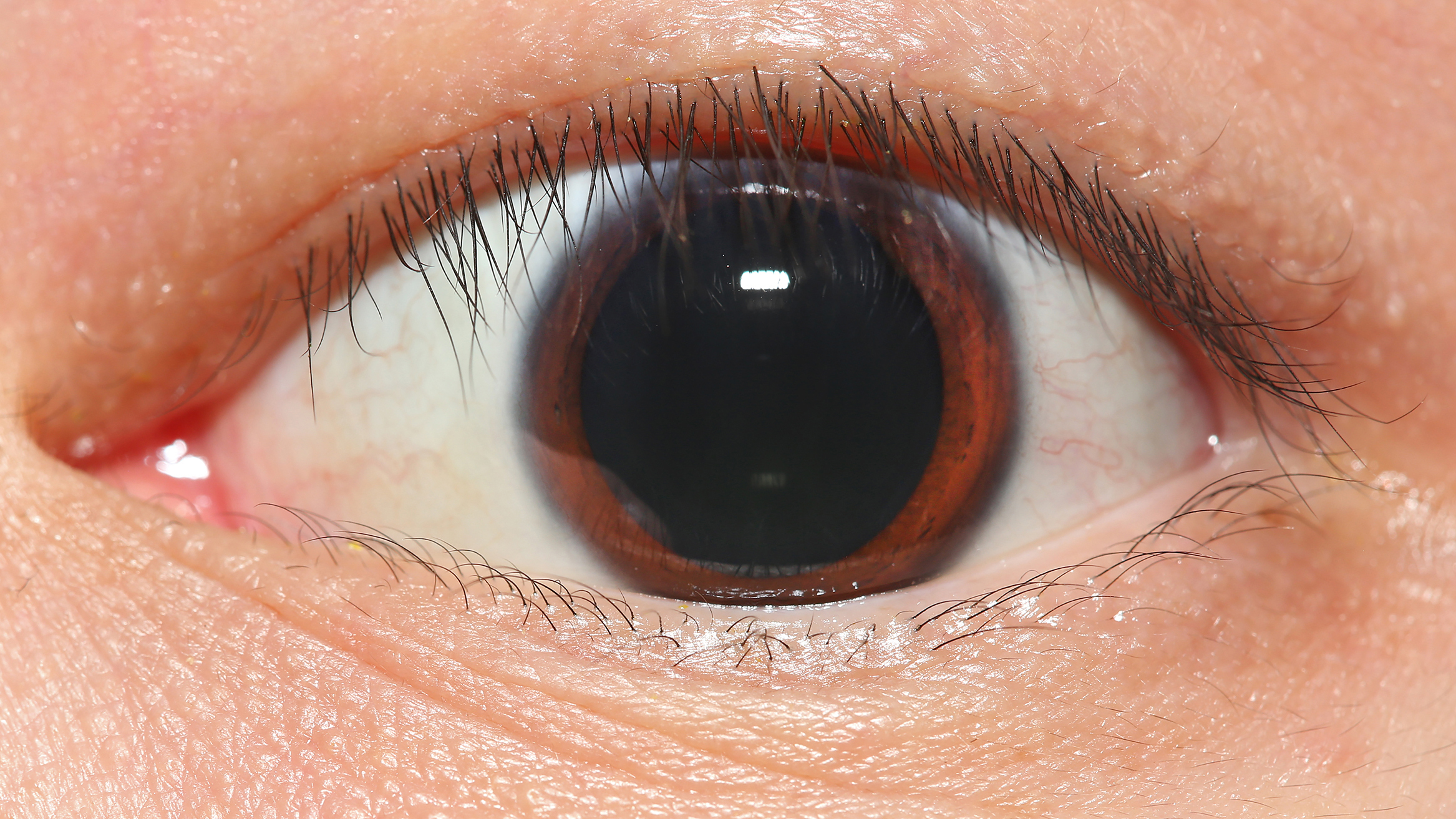
The iris dilator muscle (pupil dilator muscle, pupillary dilator, radial muscle of iris, radiating fibers), is a smooth muscle of the eye, running radially in the iris and therefore fit as a dilator. The pupillary dilator consists of a spokelike arrangement of modified contractile cells called myoepithelial cells. These cells are stimulated by the sympathetic nervous system. When stimulated, the cells contract, widening the pupil and allowing more light to enter the eye.
The ciliary muscle, pupillary sphincter muscle and pupillary dilator muscle sometimes are called intrinsic ocular muscles or intraocular muscles.
Structure
Innervation
It is innervated by the sympathetic system, which acts by releasing noradrenaline, which acts on α1-receptors. Thus, when presented with a threatening stimulus that activates the fight-or-flight response, this innervation contracts the muscle and dilates the pupil, thus temporarily letting more light reach the retina.
The dilator muscle is innervated more specifically by postganglionic sympathetic nerves arising from the superior cervical ganglion as the sympathetic root of ciliary ganglion. From there, they travel via the internal carotid artery through the carotid canal to foramen lacerum. They then enter the middle cranial fossa above foramen lacerum, travel through the cavernous sinus in the middle cranial fossa and then travel with the ophthalmic artery in the optic canal or on the ophthalmic nerve through the superior orbital fissure. From there, they travel with the nasociliary nerve and then the long ciliary nerve. They then pierce the sclera, travel between sclera and choroid to reach the iris dilator muscle. They will also pass through ciliary ganglion and travel in short ciliary nerves to reach the iris dilator muscle.
Function
The pupillary dilator acts to increase the size of the pupil to allow more light to enter the eye. It works in opposition to the pupillary constrictor. Pupil dilation occurs when there is insufficient light for the normal function of the eye, and during heightened sympathetic activity, for example in the "fight-or-flight reflex".
History
Etymology
The English name dilator pupillae muscle as currently used in the list of English equivalents of the Terminologia Anatomica, the reference-work of the official anatomic nomenclature, can be considered as a corruption of the full Latin expression musculus dilatator pupillae. The full Latin expression exhibits three words that each can be traced back to Roman antiquity. The Classical Latin name musculus is actually a diminutive of the Classical Latin name mus, and can be translated as little mouse. In the medical writings of Aulus Cornelius Celsus we can also find this specific name to refer to a muscle instead of its literal meaning. Latin musculus can be explained by the fact that a muscle looks like a little mouse that moves under the skin. In the writings of Greek philosopher Aristotle the Ancient Greek word for mouse, i.e. μῦς is also used to refer to a muscle.
Dilatator in the Latin expression musculus dilatator pupillae is derived from the classical Latin verb dilatare, to dilate, to spread out. Two possible explanations exist concerning the etymological derivation of this verb. The first explanation considers dilatare as frequentative of differere. The Latin verb differe can mean, to carry different ways, to spread abroad, to scatter, but also to delay. The other explanation considers dilatare as a compound from di- and latus, with the latter word meaning, broad or wide, hence the German name Erweiterer for Latin dilatator.
The expression dilator pupillae muscle, as used in the list of English equivalents of the Terminologia Anatomica, is actually partly Latin, i.e. dilator pupillae, with pupillae (=of the pupil), a noun in the genitive case modifying dilator, a noun in the nominative case, and partly English, i.e. muscle. In previous editions (Nomina Anatomica) this muscle was officially called the musculus dilator pupillae, The Nomina Anatomica as authorized in 1895 in Basel and in 1935 in Jena used the full Latin expression.
Additional images
File:Horner's Syndrome and Autonomic innervation of the eye.svg|Scheme showing sympathetic and parasympathetic innervation of the pupil and sites of lesion in a Horner's syndrome. File:Gray840.png|Sympathetic connections of the ciliary and superior cervical ganglia (red) (parasympathetic pathway in blue) File:Iris_Dilator_Muscle_012909.jpg|The iris dilator muscle fibers course radially through the iris.
References
References
- (2000). "Anatomy Tables – Eye". University of Michigan Medical School.
- (1987). "Muscarinic and nicotinic synaptic activation of the developing chicken iris". The Journal of Neuroscience.
- Saladin, Kenneth. (2012). "Anatomy and Physiology". McGraw-Hill.
- (March 2015). "Human ocular anatomy". Clinics in Dermatology.
- (2024). "StatPearls". StatPearls Publishing.
- Rang, H. P.. (2003). "Pharmacology". Churchill Livingstone.
- "Reflexes of the Eye". Cleveland Clinic.
- (9 December 2019). "What Happens to Your Body During the Fight-or-Flight Response?". Cleveland Clinic.
- Federative Committee on Anatomical Terminology (FCAT) (1998). ''Terminologia Anatomica''. Stuttgart: Thieme
- (2008). "Anatomical terminology and nomenclature: Past, present and highlights". Surgical and Radiologic Anatomy.
- (2001). "On the new anatomical nomenclature". Annals of Anatomy - Anatomischer Anzeiger.
- His (1895). ''Die anatomische Nomenclatur. Nomina Anatomica. Der von der Anatomischen Gesellschaft auf ihrer IX. Versammlung in Basel angenommenen Namen''. Leipzig: Verlag von Veit & Comp.{{page needed. (May 2015)
- Lewis, C.T. & Short, C. (1879). ''A Latin dictionary founded on Andrews' edition of Freund's Latin dictionary''. Oxford: Clarendon Press.{{page needed. (May 2015)
- Kraus, L.A. (1844). ''Kritisch-etymologisches medicinisches Lexikon'' (Dritte Auflage). Göttingen: Verlag der Deuerlich- und Dieterichschen Buchhandlung.{{page needed. (May 2015)
- Liddell, H.G. & Scott, R. (1940). ''A Greek-English Lexicon. revised and augmented throughout by Sir Henry Stuart Jones. with the assistance of. Roderick McKenzie.'' Oxford: Clarendon Press.{{page needed. (May 2015)
- Foster, F.D. (1891–1893). ''An illustrated medical dictionary. Being a dictionary of the technical terms used by writers on medicine and the collateral sciences, in the Latin, English, French, and German languages.'' New York: D. Appleton and Company.{{page needed. (May 2015)
- Donáth, T. & Crawford, G.C.N. (1969). ''Anatomical dictionary with nomenclature and explanatory notes.'' Oxford/London/Edinburgh/New York/Toronto/Sydney/Paris/Braunschweig: Pergamon Press.{{page needed. (May 2015)
- International Anatomical Nomenclature Committee (1966). ''Nomina Anatomica''. Amsterdam: Excerpta Medica Foundation.{{page needed. (May 2015)
- International Anatomical Nomenclature Committee (1977). ''Nomina Anatomica, together with Nomina Histologica and Nomina Embryologica''. Amsterdam-Oxford: Excerpta Medica.{{page needed. (May 2015)
- International Anatomical Nomenclature Committee (1983). ''Nomina Anatomica, together with Nomina Histologica and Nomina Embryologica''. Baltimore/London: Williams & Wilkins{{page needed. (May 2015)
- International Anatomical Nomenclature Committee (1989). ''Nomina Anatomica, together with Nomina Histologica and Nomina Embryologica''. Edinburgh: Churchill Livingstone.{{page needed. (May 2015)
- Kopsch, F. (1941). ''Die Nomina anatomica des Jahres 1895 (B.N.A.) nach der Buchstabenreihe geordnet und gegenübergestellt den Nomina anatomica des Jahres 1935 (I.N.A.)'' (3. Auflage). Leipzig: Georg Thieme Verlag.{{page needed. (May 2015)
- Stieve, H. (1949). ''Nomina Anatomica. Zusammengestellt von der im Jahre 1923 gewählten Nomenklatur-Kommission, unter Berücksichtigung der Vorschläge der Mitglieder der Anatomischen Gesellschaft, der Anatomical Society of Great Britain and Ireland, sowie der American Association of Anatomists, überprüft und durch Beschluß der Anatomischen Gesellschaft auf der Tagung in Jena 1935 endgültig angenommen.'' (4th ed.). Jena: Verlag Gustav Fischer.{{page needed. (May 2015)
This article was imported from Wikipedia and is available under the Creative Commons Attribution-ShareAlike 4.0 License. Content has been adapted to SurfDoc format. Original contributors can be found on the article history page.
Ask Mako anything about Iris dilator muscle — get instant answers, deeper analysis, and related topics.
Research with MakoFree with your Surf account
Create a free account to save articles, ask Mako questions, and organize your research.
Sign up freeThis content may have been generated or modified by AI. CloudSurf Software LLC is not responsible for the accuracy, completeness, or reliability of AI-generated content. Always verify important information from primary sources.
Report