From Surf Wiki (app.surf) — the open knowledge base
Dioctophyme renale
Species of roundworm

Species of roundworm
Dioctophyme renale, commonly referred to as the giant kidney worm, is a parasitic nematode (roundworm) whose mature form is found in the kidneys of mammals. D. renale is distributed worldwide, but is less common in Africa and Oceania. It affects fish-eating mammals, particularly mink and dogs. Human infestation is rare, but results in kidney destruction, usually of one kidney and hence not fatal. A 2019 review listed a total of 37 known human cases of dioctophymiasis in 10 countries with the highest number (22) in China. Upon diagnosis through tissue sampling, the only treatment is surgical excision.
Synonyms
Dioctophymosis, dioctophymiasis, giant kidney worm, kidney worm infection, Dioctophyme renale infection
History of discovery
Dioctophyme renale was discovered in 1583. Almost two centuries later, in 1782, Johann Goeze first described D. renale upon discovering the worms in a dog kidney. The family Dioctophymidae has only one genus (Dioctophyme), and the name of the genus was in contention (with the possibility of being Dioctophyma) for two hundred years. The issue was finally resolved by the International Commission on Zoological Nomenclature in 1987. In 2003, D. renale eggs were discovered in six human coprolites in the Neolithic site Arbon-Bleiche 3, Switzerland. This location is near a lake, which likely provided early humans with access to freshwater fish and frogs. The samples were dated from 3384 to 3370 BC, and is evidence that the prevalence of this infection was higher in early human history (before full understanding of proper cooking techniques). Eggs were also found in 2019 in a well-preserved largely fish-eating settlement in England dating to 900 BC.
Dioctophymosis
Clinical presentation in humans
Individuals with Dioctophyme renale infestation (known as dioctophymosis) typically present with unspecific symptoms including hematuria (blood in urine), nephritis, loin pain, renal enlargement, and/or renal colic (intermittent pain in the kidney area), which may result from the rare migration of worms through ureters. In some cases the fibrosis occurring after parasite infection is an incidental finding in ultrasound or CT scan, mimicking renal cancer, leading to radical nephrectomy.
Adult worms typically only infect one kidney. The kidney is destroyed because of fibrosis, the development of excess fibrous connective tissue. Global renal dysfunction is typically limited because the non-infected kidney is usually capable of assuming the increased work. However, parenchymal inflammation can lead to death in extreme circumstances.
Transmission and life cycle
Adult Dioctophyme renale inhabit the kidney (typically the right kidney). Females produce eggs which are passed in urine. In aquatic environments, eggs embryonate after 15–100 days. These eggs are ingested by an aquatic oligochaete, hatch, penetrate blood vessels, and develop into a stage three larvae. A paratenic host may then ingest the oligochaete. The oligochaete or paratenic host is then eaten by a definitive host, wherein juveniles penetrate intestinal lining and migrate to the liver. After maturing for approximately 50 days, the juveniles then migrate to the kidneys (typically the right kidney). Upon maturation, D. renale can survive for up to five years.
Definitive hosts are carnivorous mammals, notably mink, but also includes wolves, coyotes, foxes, dogs, raccoons, and weasels.
Transmission to humans typically occurs upon ingestion of raw or undercooked freshwater fish or frog.
There are multiple reservoirs for D. renale. Confirmed cases of infection have occurred in minks, dogs, swine, bears, oxen, and humans.
Morphology
D. renale is the largest nematode known to parasitize humans. Adult male worms are 20–40 cm long and 5–6 mm wide; females can grow to 103 cm in length with a width of 10–12 mm. Both sexes appear bright red in color and taper at both the anterior and posterior ends. Male D. renale worms have a bursa, which is used to attach to facilitate mating.
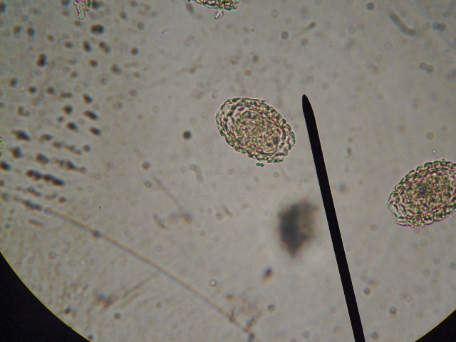
Eggs are 60–80 micrometres x 39–47 micrometres, contain an embryo, and have characteristic sculpturing of the shell. They have an oval-shape and brownish-yellow hue. Eggs have a thick shell, and the surface appears to be pitted except at the poles.
Diagnosis
The only means of obtaining a definitive diagnosis is through the identification of D. renale eggs in a patient's urine. However, obtaining patient history (i.e., if the patient has consumed undercooked or raw freshwater fish) is an important first step that can be coupled with radiological exams to search for enlarged or calcified kidneys. Urinalysis will likely show hematuria; blood tests may reveal eosinophilia.
Management
Likely because of the rarity of human cases, there is no standard treatment for D. renale infection in humans. The only known means is surgical excision of either adult worms or the infected kidney. Nephrectomy is generally considered extreme for human cases.
A physician reportedly used Ivermectin to treat a patient, who was effectively cured. The use of anti-helminth drugs has not yet been evaluated as the proper course of action to treat this infection.
Epidemiology
Though D. renale is distributed worldwide, it is markedly less frequent in Africa and Oceania, where human infection is extremely rare. Regions around the Caspian Sea have the highest number of cases, with the most occurring in Iran. Infections are also most commonly found in areas where freshwater fish is a dietary mainstay.
Non-human infections are more common worldwide, especially in areas of temperate climate. Prevalence in mink populations may be high, such as portions of Ontario or Minnesota. Similarly, some minnow populations may be as high as 50%.
Public health and prevention strategies
No public health measures have been undertaken or vaccines developed because of the rarity of human infection. The majority of D. renale infections have resulted from undercooked or raw freshwater fish consumption. Thus, the simple practice of thoroughly cooking fish prior to consumption could be promoted and lead to eradication of D. renale infection in humans.
References
References
- (5 February 2001). "Animal Parasitology". Kansas State University.
- (22 February 2009). "Urinary System Diseases, Animals". Parasitology Research & Encyclopedic Reference of Parasitology.
- (22 February 2009). "Homo sapiens diseases - Metazoa". Molecular Medicine.
- (22 February 2009). "Kidney Worm: Dioctophymiasis and Eustrongylidiasis". Tropical Medicine Central Resource International Society of Radiology.
- (2019). "A human case of ''Dioctophyma renale'' (giant kidney worm) accompanied by renal cancer and a retrospective study of dioctophymiasis". Parasite.
- (22 February 2009). "Dioctophyme renale Infection in Bears (Parasitic Disease Summary)".
- (22 February 2009). "An Introduction to FOODBORNE DISEASES & HACCP Systems". Mediterranean Zoonoses Control Center/World Health Organization.
- (22 February 2009). "Infectious Diseases". Gideon.
- [https://archive.org/details/biostor-5104 ICZN Opinion 1552]
- (October 2003). "Dioctophymidae Eggs in Coprolites From Neolithic Site of Arbon-Bleiche 3 (Switzerland)". [[Journal of Parasitology]].
- Ian Sample. (16 August 2019). "Bronze age meals in the marshes – seasoned with parasitic worms". The Guardian.
- (October 2013). "Case Report: A rare case of a 39-year-old male with a parasite called Dioctophyma renale mimicking renal cancer at the computed tomography of the right kidney". Parasitology International.
- (January 1985). "Centrachid Fish as Paratenic Hosts of the Giant Kidney Worm, Dioctophyma Renale (Goeze, 1782), in Ontario, Canada". Journal of Wildlife Diseases.
- "Giant Kidney Worm (Dioctophyme renale) in Dogs". PetEducation.com.
- Mace, T. F.. (January 1976). "Lessions in Mink (Mustela vision) Infected with Giant Kidney Worm (Dioctophyma renale)". Journal of Wildlife Diseases.
- (April 2009). "Dioctophyme renale (Nematoda, Dioctophymatidae) Infection in the Crab-eating Fox (Cerdocyon thous) from Brazil". Journal of Wildlife Diseases.
- (April 2007). "Giant kidney worm (Dioctophyma renale) infections in dogs from Northern Paraná, Brazil". Veterinary Parasitology.
- (January 2001). "Prevalence of Giant Kidney Worm (Dioctophyma renale) in Wild Mink (Mustela vison) in Minnesota". [[American Midland Naturalist]].
- (October 1990). "Dioctophymosis in the Little Grison (Galictis cuja)". Journal of Wildlife Diseases.
- (January 2003). "Infestation of the Human Kidney with Dioctophyma renale". Urologia Internationalis.
This article was imported from Wikipedia and is available under the Creative Commons Attribution-ShareAlike 4.0 License. Content has been adapted to SurfDoc format. Original contributors can be found on the article history page.
Ask Mako anything about Dioctophyme renale — get instant answers, deeper analysis, and related topics.
Research with MakoFree with your Surf account
Create a free account to save articles, ask Mako questions, and organize your research.
Sign up freeThis content may have been generated or modified by AI. CloudSurf Software LLC is not responsible for the accuracy, completeness, or reliability of AI-generated content. Always verify important information from primary sources.
Report