From Surf Wiki (app.surf) — the open knowledge base
Central canal
Cerebrospinal fluid-filled space around the spinal cord
Cerebrospinal fluid-filled space around the spinal cord
| Field | Value |
|---|---|
| Name | Central canal of spinal cord |
| Latin | canalis centralis medullae spinalis |
| Image | Medulla spinalis - Section - English.svg |
| Caption | Cross-section through cervical spinal cord. |
| Image2 | Medulla spinalis - Substantia grisea - English.svg |
| Location | Centre of the spinal cord |
The central canal (also known as spinal foramen or ependymal canal) is the cerebrospinal fluid-filled space that runs through the spinal cord. The central canal lies below and is connected to the ventricular system of the brain, from which it receives cerebrospinal fluid, and shares the same ependymal lining. The central canal helps to transport nutrients to the spinal cord as well as protect it by cushioning the impact of a force when the spine is affected.
The central canal represents the adult remainder of the central cavity of the neural tube. It generally occludes (closes off) with age.
Structure
The central canal below at the ventricular system of the brain, beginning at a region called the obex where the fourth ventricle, a cavity present in the brainstem, narrows.
The central canal is located in the third of the spinal cord in the cervical and thoracic regions. In the lumbar spine it enlarges and is located more centrally. At the conus medullaris, where the spinal cord tapers, it is located more .
Terminal ventricle
The terminal ventricle (ventriculus terminalis, fifth ventricle or ampulla caudalis) is the widest part of the central canal of the spinal cord that is located at or near the conus medullaris. It was described by Stilling in 1859 and Krause in 1875. Krause introduced the term fifth ventricle after observation of normal ependymal cells. Although the terminal ventricle is visible in the fetus and children, it is usually absent in adults.
Sometimes, the terminal ventricle is observed by MRI or ultrasound in children less than 5 years old.
Microanatomy
Central gelatinous substance The central canal shares the same ependymal lining as the ventricular system of the brain.
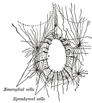
The canal is lined by ciliated, column-shaped cells, outside of which is a band of gelatinous substance, called the substantia gelatinosa of Rolando also substantia gelatinosa centralis or central gelatinous substance of spinal cord. This gelatinous substance consists mainly of neuroglia, but contains a few nerve cells and fibers; it is traversed by processes from the deep ends of the columnar ciliated cells which line the central canal.
Development
The central canal represents the adult remainder of the central cavity of the neural tube. It generally occludes (closes off) with age.
Function
The central canal carries cerebrospinal fluid (CSF), which it receives from the ventricular system of the brain. The central canal helps to transport nutrients to the spinal cord as well as protect it by cushioning the impact of a force when the spine is affected.
Clinical significance
Syringomyelia is a disease caused by the blockage of the central canal. Blockage of the central canal usually occurs at the lower cervical and upper thoracic levels. This typically damages white matter fibers that cross in anterior white commissure, leading to the loss of temperature, pain, and motor function at the affected levels on side opposite to the damage.
Other relevant conditions include:
- Spina bifida
- Arnold-Chiari syndrome
- Spinal tumor
- Myelomeningocele
- Syringomyelia
- Hydromyelia. In hydromyelia, a dilation of the central canal of the spinal cord is caused by an increase of cerebrospinal fluid.
- Syringohydromyelia (i.e., both Syringomyelia and Hydromyelia)
- Tethered cord
In some cases, the terminal ventricle may cause clinical symptoms due to its expansion.
References
Tomsick T, Peak E, Wang L: Fluid-Signal Structures in the Cervical Spinal Cord on MRI: Anterior Median Fissure vs. Central Canal. AJNR 2017; 38:840–45
Tomsick T, Wang L, Zuccarello M, Ringer AJ. Fluid-signal structures in the cervical spinal cord on MRI in Chiari patients: Central canal or anterior median fissure? AJNR Am J Neuroradiol. 2021 Apr;42(4):801-806. doi: 10.3174/ajnr.A7046. Epub 2021 Mar 11.PMID: 33707286
References
- (2016). "The Human Central Canal of the Spinal Cord: A Comprehensive Review of its Anatomy, Embryology, Molecular Development, Variants, and Pathology". Cureus.
- (1999). "Age-related morphologic changes of the central canal of the human spinal cord". Acta Neuropathol..
- (August 2024). "ventriculus terminalis".
- (June 2005). "Fifth ventricle: an unusual cystic lesion of the conus medullaris". Spinal Cord.
- (1989). "Gray's Anatomy".
- (July 2008). "ventriculus terminalis".
- (March 2002). "Cyst of the medullary conus: malformative persistence of terminal ventricle or compressive dilatation?". Neurosurgical Review.
- (2018-04-25). "imaging in syringohydromyelia".
This article was imported from Wikipedia and is available under the Creative Commons Attribution-ShareAlike 4.0 License. Content has been adapted to SurfDoc format. Original contributors can be found on the article history page.
Ask Mako anything about Central canal — get instant answers, deeper analysis, and related topics.
Research with MakoFree with your Surf account
Create a free account to save articles, ask Mako questions, and organize your research.
Sign up freeThis content may have been generated or modified by AI. CloudSurf Software LLC is not responsible for the accuracy, completeness, or reliability of AI-generated content. Always verify important information from primary sources.
Report